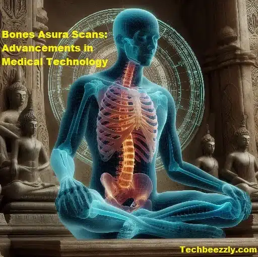
Bones asura scans health is essential for overall well-being, yet it’s often overlooked until problems arise. Fortunately, advancements in medical technology have made it easier to assess. The monitor bone health through procedures like bones asura scans.
What are Bone Asura Scans?
Bones asura scans, also known as bone density scans or DXA scans (Dual-energy X-ray absorptiometry). They have specialized imaging tests that measure bone mineral density. They have commonly used to diagnose osteoporosis, a condition characterized by weakened and fragile bones.
How do Bone Asura Scans work?
During a bones asura scans, the patient lies on a table while a machine passes over the body, emitting low-dose X-rays. These X-rays have absorbed differently by bone and soft tissue, allowing the technician to measure bone density accurately. The results have typically expressed as T-scores and Z-scores. Which compare the patient’s bone density to that of a healthy young adult.
Early detection of bone diseases:
One of the primary benefits of bones asura scans is their ability to detect bone loss at an early stage, even before symptoms occur. This early detection enables timely intervention and treatment to prevent fractures and other complications.
Monitoring bone health:
For individuals already diagnosed with osteoporosis or at risk for bone-related issues. It regular bones asura scans are essential for monitoring bone density changes over time. This information helps healthcare providers adjust treatment plans as needed to maintain optimal bone health.
Preparation for Bone Asura Scans
Before undergoing a bones asura scans, patients have typically advised to wear loose, comfortable clothing and avoid wearing any metal objects that could interfere with the imaging process. There are usually no dietary restrictions, and the procedure itself is quick and painless, taking only a few minutes to complete.
Accurate assessment of bone density
bones asura scans provide a precise measurement of bone mineral density, allowing healthcare providers to assess the risk of fractures accurately. This information helps guide treatment decisions and lifestyle recommendations to improve bone health.
Non-invasive and painless procedure
Unlike some other imaging techniques, such as bone biopsies, bones asura scans are non-invasive and painless. Patients simply lie on a table while the scanner passes overhead, making it a comfortable experience for most individuals.
Risks and Limitations of Bone Asura Scans
Although the amount of radiation used in bones asura scans is minimal, some individuals may have concerns about exposure. However, the benefits of early detection and monitoring of bone health often outweigh the risks associated with radiation exposure.
Factors that may affect scan accuracy
Certain factors, such as obesity or the presence of metal implants, can affect the accuracy of bones asura scans results. It’s essential to inform the healthcare provider of any relevant medical history or conditions before undergoing the procedure to ensure accurate results.
Understanding T-scores and Z-scores
T-scores compare bone density to that of a healthy young adult of the same gender, while Z-scores compare bone density to that of an individual’s age group. A T-score of -1.0 or above is considered normal, while a T-score between -1.0 and -2.5 indicates low bone mass (osteopenia), and a T-score of -2.5 or lower indicates osteoporosis.
Abnormal bones asura scans results may indicate a higher risk of fractures and other bone-related issues. Depending on the severity of the findings, healthcare providers may recommend lifestyle changes, medication, or other interventions to improve bone health and reduce fracture risk.
Comparing Bone Asura Scans with other imaging techniques
While X-rays can detect fractures and abnormalities in bone structure, they do not provide information about bone density. MRIs, on the other hand, can offer detailed images of soft tissue but are less effective at assessing bone density compared to bones asura scans.
When each imaging technique is preferred
bones asura scans have typically preferred for assessing bone density and diagnosing conditions like osteoporosis. X-rays and MRIs may used in conjunction with bones asura scans to provide a comprehensive evaluation of bone health and identify any additional issues.
Who should consider Bone Asura Scans?
Women over the age of 65 and men over the age of 70 are at an increased risk of osteoporosis and may benefit from bones asura scans. Additionally, individuals with a family history of osteoporosis, low body weight. Or certain medical conditions may also advised to undergo screening.
Those with a history of fractures or bone-related issues
Individuals who have experienced fractures or have conditions that affect bone health. Such as rheumatoid arthritis or hyperparathyroidism, may benefit from bones asura scans to assess their bone density and monitor changes over time.
Many insurance plans cover bones asura scans for individuals at risk of osteoporosis or those who have already been diagnosed